Lateral Calcaneal Slide Osteotomy

What is a calcaneal osteotomy.
Lateral calcaneal slide osteotomy. Patients with stage 2b flatfoot have lateral pain due to calcaneofibular impingement and a more severely abducted forefoot. Arthrex s recently launched minimally invasive surgery platform allows surgeons to perform this osteotomy through a tiny incision. By extending the length of the calcaneus at the location of the talonavicular joint the talonavicular joint can be rotated from an abducted to neutral alignment. These patients benefit from posterior tibial tendon repair or flexor digitorum longus transfer along with a medial calcaneal slide osteotomy.
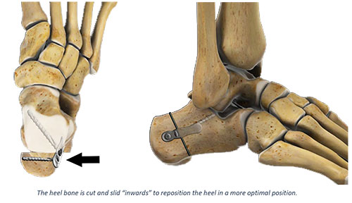
The posterior osteotomy fragment is manually mobilized and shifted laterally. Surgeons commonly perform the medial slide calcaneal osteotomy in conjunction with a flexor digitorum longus tendon transfer lateral column lengthening or a medial cuneiform osteotomy or arthrodesis. The surgeon also needs to address any equinus deformity associated with the pathology. Surgeons can then achieve fixation using the 7 0 mm compression ft screws or the 6 5 compression pt screws.
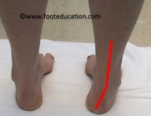
The incision was carried down through the skin only with a 15 blade knife. The osteotomy is performed through an oscillating saw. Dr performed a lateral slding calcaneal osteotomy along with a lateral column lengthening need help with cpt code. When the heel is observed from behind it is generally situated in line with the leg.
Use a sagittal saw or osteotome to perform the calcaneus osteotomy this is an osteotomy from proximal lateral to distal medial that starts 2 2 5 cm proximal to the cc joint and exits between the anterior and middle facets this is a complete osteotomy through the medial cortex. The calcaneus or heel bone plays an important role in walking. A calcaneal osteotomy is a common technique used to treat stage ii flatfoot deformity. Anterior calcaneal osteotomy lateral column lengthening a lateral column lengthening is performed typically to correct the forefoot abduction aspect of the deformity.
If needed a laterally based wedge can be removed and or the osteotomy fragment can be translated cranially. A specialized calcaneal slide plate was an effective fixation device for both medial and lateral calcaneal slide osteotomies for a variety of foot and ankle conditions. The union rate was 100 and none of the patients had hardware symptoms which is an improvement on published reports of symptomatic hardware after calcaneal slide osteotomy. The heel bone called the calcaneus is the main bone that lies in the heel of the hindfoot.
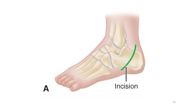
The lateral calcaneus cortex is exposed using a lateral incision. A calcaneal osteotomy comprises of making a cut across the heel bone and shifting it toward the inside medial or outside lateral.

Medial Calcaneal Sliding Osteotomy
Http Citeseerx Ist Psu Edu Viewdoc Download Doi 10 1 1 1026 8013 Rep Rep1 Type Pdf

Calcaneal Sliding Osteotomy With Lateral Plate Fixation Resurgens Orthopaedics

Wheeless Textbook Of Orthopaedics

Congenital And Acquired Neurologic Disorders Musculoskeletal Key

Medial Calcaneal Sliding Osteotomy Surgery In Washington D C Maryland And Northern Virginia

Adult Acquired Flat Foot Pes Planus Deformity Orthopaedic Product News
Http Fai Sagepub Com Content 35 3 238 Full Pdf Html

Figure 3 From What Is The Role And Limit Of Calcaneal Osteotomy In The Cavovarus Foot Semantic Scholar

Calcaneal Sliding Osteotomy Stryker
Tibial Nerve Palsy Associated With Lateralizing Calcaneal Osteotomy Case Reviews And Technical Tip Semantic Scholar

Figure 1 From Preoperative Planning And Intraoperative Technique For Accurate Realignment Of The Dwyer Calcaneal Osteotomy Semantic Scholar

Ankle Fusion Chicopee Arthroscopic Ankle Cartilage Repair Springfield East Longmeadow

Figure 2 From Calcaneus Osteotomy Semantic Scholar
Flat Foot Anatomy And Current Treatment Ppt Download

Arthrex Calcaneus Step Plate
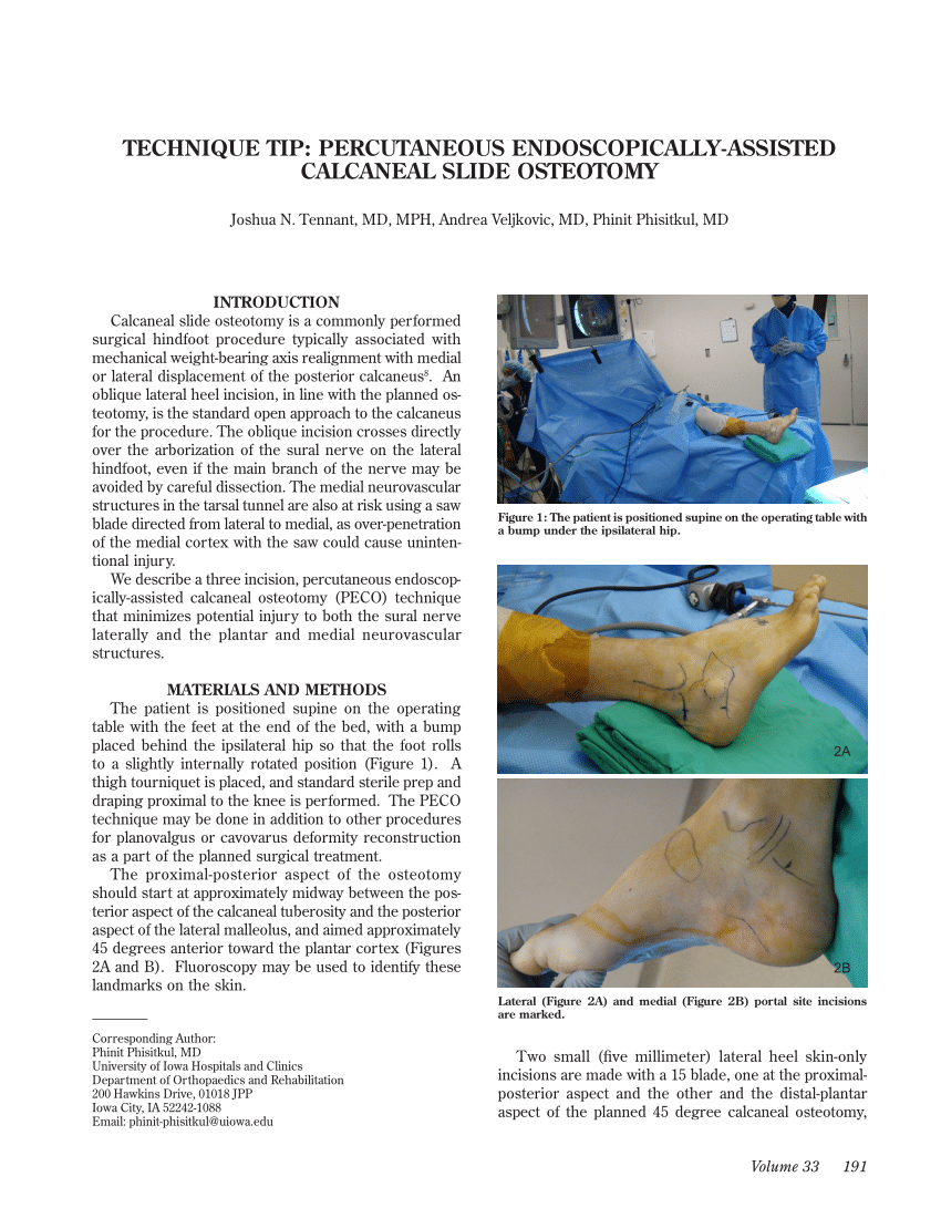
Pdf Technique Tip Percutaneous Endoscopically Assisted Calcaneal Slide Osteotomy
The Foot In Cp Part 2 Of 3

Left Ankle Reconstruction Calcaneal Osteotomy Medical Illustration Human Anatomy Drawing Anatomy Illustration

The Photograph A And Standing Lateral Radiograph B Of Left Foot Download Scientific Diagram
Painful Flatfoot Guide
Calcaneal Osteotomy Footeducation

Evans Lateral Column Lengthening And Cotton Osteotomy Musculoskeletal Key

Pdf Calcaneal Osteotomy Safe Zone To Prevent Neurological Damage Fact Or Fiction
Https Encrypted Tbn0 Gstatic Com Images Q Tbn 3aand9gcrzcauc5ccpdxgi1ophbi6ez Sewcgsaukt6anxsmfg2gb1gjvt Usqp Cau

Paragon28 Calc Slide Plate Orthopedic Implants Paragon28

Key Pearls Of Calcaneal Osteotomies Podiatry Today

Normal Appearance Of Subtalar Arthroereisis A Lateral And B Download Scientific Diagram
Http Www Apma Org Files Apma 20flatfoot 20reconstruction Pdf

Management Of Symptomatic Flexible Flatfoot In Adolescents By Mosca S Lateral Calcaneal Lengthening

Pdf Calcaneal Displacement Osteotomies Less Soft Tissue Irritation In Lateral Compression Plate Than Screws Semantic Scholar

Arthrex Calcaneal Osteotomy For Flatfoot

Figure 2 From Geometry Of The Lateral Sliding Closing Wedge Calcaneal Osteotomy Semantic Scholar

Lateral Column Lengthening Footeducation

Figure 1 From Minimally Invasive Calcaneal Osteotomy Does The Shannon Burr Endanger The Neurovascular Structures A Cadaveric Study Semantic Scholar

Surgical Management Of Calcaneal Malunion Sciencedirect

Medial Displacement Calcaneal Osteotomy Youtube
Lecture 42 Shah Calcaneal Malunions

Medial Sliding Osteotomy Of The Calcaneus A 59 Year Old Male With Download Scientific Diagram

Pdf A Mid Term Follow Up Of Koutsogiannis Osteotomy In Adult Acquired Flatfoot Stage Ii And Early Stage Iii
Https Www Med Upenn Edu Orthoresidents Assets User Content Documents Oite Fa Review Pdf

Comparison Of Lateral Opening Wedge Calcaneal Osteotomy And Medial Calcaneal Sliding Opening Wedge Cuboid Closing Wedge Cuneiform Osteotomy For Correction Of Planovalgus Foot Deformity In Children Semantic Scholar
Https Journals Sagepub Com Doi Pdf 10 1177 1071100717728678
Mce 2016 Semester Ii Foot Deformities Benha University Orthopaedi
Metatarsal And Calcaneal Osteotomies For Cavus Foot Musculoskeletal Key

Normal Appearance Of Medial Displacement Calcaneal Osteotomy A Download Scientific Diagram

Jaypeedigital Ebook Reader

Pdf A Prospective Study Of Japas Osteotomy In Paralytic Pes Cavus Deformity In Adolescent Feet
